In Vitro to In Vivo Extrapolation (QIVIVE) for Predicting Drug-Induced Liver Toxicity
2024, EUROTOX
DOWNLOAD
View Details
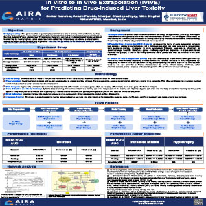
In Vitro to In Vivo Extrapolation (QIVIVE) for Predicting Drug-Induced Liver Toxicity
2024, EUROTOX
The objective of this study was to create a model for predicting drug-induced liver toxicity by identifying crucial genes linked to cell toxicity in in vitro settings and assessing their ability to predict toxicity in in vivo. The objective was to establish a connection between in vitro and in vivo data by employing quantitative in vitro to in vivo extrapolation (QIVIVE) techniques, which have the potential to reduce animal sacrifice in in vivo studies.
Machine Learning for Rodent Liver Toxicity Prediction: Leveraging Drug Substructure Properties
2024, EUROTOX
DOWNLOAD
View Details
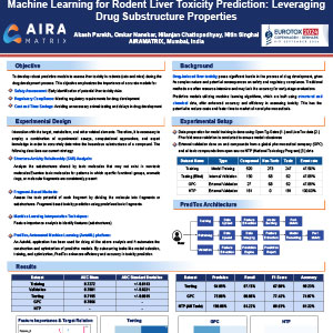
Machine Learning for Rodent Liver Toxicity Prediction: Leveraging Drug Substructure Properties
2024, EUROTOX
Assessing liver toxicity in rodents (rats, mice) is crucial in various drug development processes and is important for safety assessment, regulatory compliance, cost, and time savings for pharmaceutical companies, necessitating robust predictive models. This study showcases the efficacy of machine learning in predicting rodent liver toxicity, offering a potential solution for compound-level toxicity assessments and informed drug development strategies.
Deep Learning-based Method for Anatomical Subsite-wise Evaluation of Single Cell Necrosis and Vacuolation of Neuron/Nerve Fiber of CNS Toxicity in Rats
2024, STP Annual Symposium
DOWNLOAD
View Details
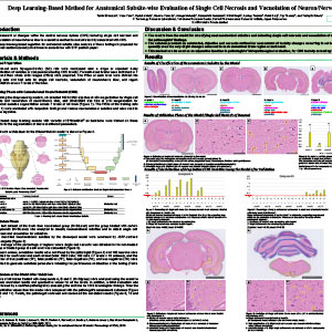
Deep Learning-based Method for Anatomical Subsite-wise Evaluation of Single Cell Necrosis and Vacuolation of Neuron/Nerve Fiber of CNS Toxicity in Rats
2024, STP Annual Symposium
Assessment of changes within the central nervous system (CNS) including single cell necrosis and vacuolation of neuron/nerve fiber is a sensitive method to assess toxicity. A deep learning-based algorithm for analysis of single-cell necrosis and vacuolation is proposed for 7 levels of rat brain.
Deep Learning Solution for the Automated Assessment of the Rodent Thymus
2024, STP Annual Symposium
DOWNLOAD
View Details
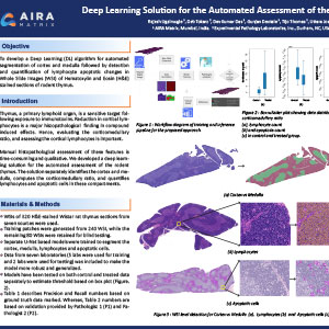
Deep Learning Solution for the Automated Assessment of the Rodent Thymus
2024, STP Annual Symposium
The thymus, being a primary lymphoid organ, is a sensitive target following exposure to immunotoxins. Reduction in cortical lymphocytes is an important histopathological finding in compound-induced effects. Hence evaluating the cortico-medullary ratio, and assessing the cortical lymphocytes is important. Manual histopathological assessment of these features can be a time-consuming process with subjective outputs. We developed a deep-learning solution for the automated assessment of the rodent thymus. The solution separately identifies the cortex and medulla, computes the cortico-medullary ratio, and quantifies lymphocytes and apoptotic cells in each compartment.
Solving the Reference Pathologist Paradox in Machine Learning Development for Histopathology Scoring
2024, STP Annual Symposium
DOWNLOAD
View Details
Solving the Reference Pathologist Paradox in Machine Learning Development for Histopathology Scoring
2024, STP Annual Symposium
Development of machine learning (ML) algorithms for scoring histology slides for nonclinical toxicology commonly involves training against example histopathology findings. This approach creates an ML performance boundary based on the list of diagnoses included in training and the sensitivity of the pathologist who made the diagnoses. We hypothesized that a ML development strategy that does not require training against histopathology findings could increase algorithm sensitivity.
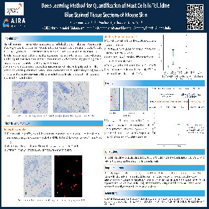
Deep Learning Method for Quantification of Mast Cells in Toluidine Blue-Stained Tissue Sections of Mouse Skin
2023, STP Annual Symposium
DOWNLOAD
View Details
Deep Learning Method for Quantification of Mast Cells in Toluidine Blue-Stained Tissue Sections of Mouse Skin
2023, STP Annual Symposium
Mast cells play a significant role in skin immunity and the pathogenesis of multiple skin diseases, including atopic dermatitis, scleroderma, contact dermatitis, blistering cutaneous disorders, and chronic graft versus host disease. It is also responsible for physiological functions such as innate and adaptive responses, angiogenesis, regulation of vasodilation, vascular homeostasis, and detoxification of venom. A common characteristic across skin diseases is an increase in mast cells, which undergo degranulation in the affected skin. Accurate quantification of mast cells is an important step in assessing the efficacy of study models for skin diseases. We propose a deep learning (DL)-based method for quantification of mast cells in skin sections of mice stained with toluidine blue.
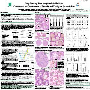
Deep Learning-Based Image Analysis Model for Evaluation of Testicular and Epididymal Lesions in Rats
2023, STP Annual Symposium
DOWNLOAD
View Details
Deep Learning-Based Image Analysis Model for Evaluation of Testicular and Epididymal Lesions in Rats
2023, STP Annual Symposium
Spermatogenic staging and assessment of testicular toxicities in rat tissue sections are time-consuming and require the expertise of well-trained pathologists. Deep Learning (DL)-based image analysis is increasingly being used for preclinical safety-assessment studies in the pharmaceutical industry. Here we present a DL-based solution for classifying 11 stage groups of spermatogenesis namely stages I, II-III, IV-VI, VII, VIII, IX, X, XI, XII-XIII, XIV, XIV, and "Stage Not Analyzed" based on normal testicular tissue structure. In addition, the solution for identifying and quantifying testicular and epididymal lesions in rat toxicology studies is also shown.
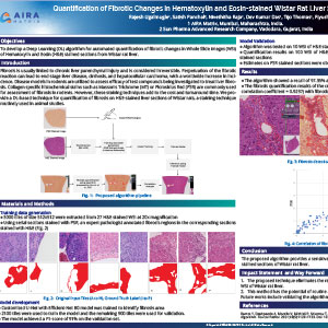
Quantification of Fibrotic Changes in Hematoxylin and Eosin-Stained Wistar Rat Liver Sections Using Deep Learning
2022, STP Annual Symposium
DOWNLOAD
View Details
Quantification of Fibrotic Changes in Hematoxylin and Eosin-Stained Wistar Rat Liver Sections Using Deep Learning
2022, STP Annual Symposium
Fibrosis is usually linked to chronic liver parenchymal injury and is considered irreversible. Perpetuation of the fibrotic reaction can lead to end-stage liver disease, cirrhosis, and hepatocellular carcinoma, with a worldwide increase in incidence. Disease models in rodents are utilized to assess the efficacy of test compounds being investigated to treat liver fibrosis. Collagen-specific histochemical stains such as Masson's Trichrome (MT) or Picrosirius Red (PSR) are commonly used for the assessment of fibrosis in rodents. However, these staining techniques add to the cost and turnaround time. We provide a DL-based technique for quantification of fibrosis on H&E-stained liver sections of Wistar rats. A staining technique routinely used in animal studies.
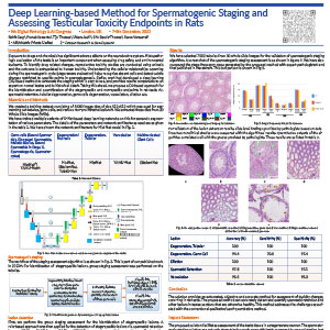
Deep Learning-Based Method for Spermatogenic Staging and Assessing Testicular Toxicity Endpoints in Rats
2022, DP & AI Congress
DOWNLOAD
View Details
Deep Learning-Based Method for Spermatogenic Staging and Assessing Testicular Toxicity Endpoints in Rats
2022, DP & AI Congress
Exposure to drugs and chemicals has significant adverse effects on the reproductive system. Histopathology evaluation of the testis is an important component when assessing drug safety and environmental toxicants. Understanding the cellular relationships occurring during the spermatogenic cycle helps recognize absent cells and detect subtle changes restricted to specific points in spermatogenesis. Earlier, we had developed a deep learning (DL)-based method to automate the staging which is easy to use and provides results comparable to an expert on normal testes and to historical data. Taking this ahead, we propose a DL-based approach for the identification and quantification of the stage-specific and non-specific endpoints in rat testis.
Automated Identification and Quantification of Pancreatic Pathology in Rodents
2022, STP Annual Symposium
DOWNLOAD
View Details
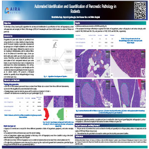
Automated Identification and Quantification of Pancreatic Pathology in Rodents
2022, STP Annual Symposium
Drug-induced pancreatic injury in preclinical toxicology studies is a serious liability in drug development. Pancreatic toxicity is generally characterized by dysregulation of lipid metabolism and edema in early reversible stages, followed by massive necrosis resulting in inflammation, with or without fibrosis at the advanced irreversible stages. Some patients with pancreatitis can also develop pancreatic cancer. Therefore, accurate identification and characterization of test compound-induced pancreatic lesions in preclinical toxicity studies is important to understand clinical translatability. Islet cell hyperplasia, acinar cell apoptosis, and atrophy are the commonly observed pathological lesions in the pancreas in rodent studies. We present a DL-based method to quantify these histopathological changes in rodent pancreas.
Deep Learning-Based Image Analysis Algorithm for Classification and Quantification of Multiple Histopathological Lesions of the Rat Liver and Kidney
2022, STP Annual Symposium
DOWNLOAD
View Details
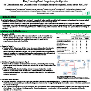
Deep Learning-Based Image Analysis Algorithm for Classification and Quantification of Multiple Histopathological Lesions of the Rat Liver and Kidney
2022, STP Annual Symposium
Artificial Intelligence (AI)-based image analysis is increasingly being used for preclinical safety-assessment studies in the pharmaceutical industry. In this study, we present a Deep Learning (DL)-based method for the classification and quantification of multiple histopathological lesions in rodent liver and kidney. The trained algorithms were validated using 255 liver Whole Slide Images (WSIs) to detect, classify, and quantify the seven findings in the liver. A modified form of the U-Net DL Model was trained using data from WSIs of 92 liver sections and 90 kidney sections. The trained model was used for identifying and quantifying 7 types of histopathology findings in both liver (vacuolation, bile duct hyperplasia, single-cell necrosis, microgranuloma, EMH, and hypertrophy) and kidney (vacuolation, basophilia/degeneration/regeneration tubule, dilation, hyaline cast, mineralization, mononuclear cell infiltration, and cyst). The algorithm was validated by comparing the results with pathologists' findings on 255 liver sections and 285 kidney sections.
Deep Learning Models for Quantifying Testicular Toxicity in Rats
2022, STP Annual Symposium
DOWNLOAD
View Details
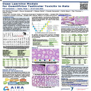
Deep Learning Models for Quantifying Testicular Toxicity in Rats
2022, STP Annual Symposium
Recognizing the 14 stages in the rat testis cycle is a difficult task. We have developed a deep learning (DL)based method to automate the staging which is easy to use and provides results comparable to an expert on normal testes, and historical data. Taking this ahead, we propose a DL-based method for identification and quantification of findings in rat testes. We have concentrated on a subset of findings from the INHAND nomenclature. We selected several Janssen toxicology studies on which early stages findings (spermatid retention after one month) and more chronic, degenerative findings were recorded. We have developed DL models to detect those findings and compared them to pathologists' results.
Deep Learning-Based Image Analysis Algorithm for Classification and Quantification of Multiple Histopathological Lesions of the Rat Liver
2022, JSTP Annual Symposium
DOWNLOAD
View Details
Deep Learning-Based Image Analysis Algorithm for Classification and Quantification of Multiple Histopathological Lesions of the Rat Liver
2022, JSTP Annual Symposium
Artificial Intelligence (AI)-based image analysis is increasingly being used for preclinical safety-assessment studies in the pharmaceutical industry. In this study, we present an AI-based solution for preclinical toxicology studies. We trained a set of algorithms to learn and quantify seven types of typical histopathological findings (including vacuolation, bile duct hyperplasia, and single cell necrosis) in whole slide images (WSIs) of the livers of young Sprague Dawley (SD) rats by using a U-Net-based deep learning network. The trained algorithms were validated using 255 liver WSIs to detect, classify, and quantify the seven findings in the liver.
Automated Method for 1) Assessing the Phases of Oestrus Cycle and 2) Quantifying Ovarian Follicles in Rat
2022, DP & AI Congress
DOWNLOAD
View Details
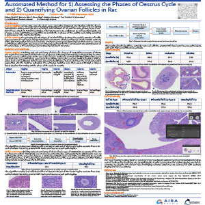
Automated Method for 1) Assessing the Phases of Oestrus Cycle and 2) Quantifying Ovarian Follicles in Rat
2022, DP & AI Congress
Oestrus Staging: Various drugs and chemicals cause endocrine disruption and interfere with the female reproductive system. Accurate and consistent determination of rat oestrus cycle phases helps understand these effects in nonclinical studies. This task needs to be performed by skilled pathologists. We propose a deep learning (DL)-based algorithm to identify the 4 phases of the oestrus cycle using the combined morphology of a rat's vagina and uterus.
Follicle Counting: The counting of early stages of ovarian follicles to study the possible effects on fertility is recommended/required in multigenerational reproductive studies performed on rats. Manual counting of different ovarian follicles is a tedious, error-prone, and time-consuming task that must be done by well-trained pathologists. We propose an automated solution for the identification and quantification of primordial type 1, primordial type 2, and growing follicles in rat ovaries.
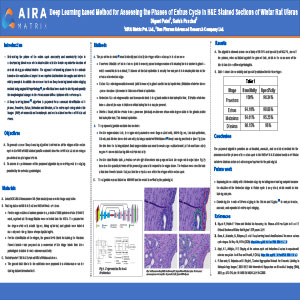
Deep Learning-Based Method for Assessing the Phases of Estrus Cycle in H&E Stained Sections of Wistar Rat Uterus
2021, STP Annual Symposium
DOWNLOAD
View Details
Deep Learning-Based Method for Assessing the Phases of Estrus Cycle in H&E Stained Sections of Wistar Rat Uterus
2021, STP Annual Symposium
Determining the phase of the estrus cycle accurately and consistently helps in understanding the interference of chemical entities with the female reproductive function of animals during pre-clinical studies. The approach of ascribing phases to the animals based on the evaluation of a synchronous system that includes the vagina and uterus is widely accepted. In addition to our novel work on Deep Learning-based estrus staging method using vaginal histopathology, an effort has been made to identify and quantify the morphological changes in the uterus across different phases of the estrus cycle. A Deep Learning-based algorithm is proposed for an accurate identification of 4 phases, Proestrus, Estrus, Metestrus, and Diestrus, of the estrus cycle using whole slide images (WSI) of conventional hematoxylin and eosin-stained sections of Wistar rat uterus.
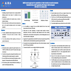
Multi-Modal Approach for Prediction of Skin Toxicity in Small Molecules
2021, EUROTOX Congress
DOWNLOAD
View Details
Multi-Modal Approach for Prediction of Skin Toxicity in Small Molecules
2021, EUROTOX Congress
Calculating precomputed descriptor values is a common method for identifying molecular data, however it is ineffective for large molecular sizes. To accurately characterize skin hazardous data, we devised a multimodal technique. We have also created a tool that reveals the class designation as well as the essential substructure found in skin-hazardous compounds.
